Types of mri Machines

1.5T is considered state-of-the-art in medical imaging. Today, the standard for MRI in a clinical setting is a 1.5T MRI. A 1.5T machine is faster than lower strength MRIs and is ideal for abdomens and chest MRIs (where patients are asked to hold their breath for the MRI sequence).

3T MRI: the newest and most powerful. The image quality of an MRI depends on signal and field strength. So a 3T machine has much more signal than a 1.5T machine.

The High-field Wide-bore MRI is considerably wider than a traditional MRI. Imagine the opening to be about the size of a hula hoop. The wider bore makes the exam a good option for claustrophobic, obese or broad-shouldered patients.
A breast MRI is mainly used for women who have been diagnosed with breast cancer, to help measure the size of the cancer, look for other tumors in the breast, and to check for tumors in the opposite breast. For certain women at high risk for breast cancer, a screening MRI is recommended along with a yearly mammogram.

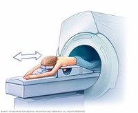
What is Cardiac MRI? Magnetic resonance imaging (MRI) is a noninvasive medical test that physicians use to diagnose medical conditions. MRI uses a powerful magnetic field, radio frequency pulses and a computer to produce detailed pictures of organs, soft tissues, bone and virtually all other internal body structures.

Provision for Alternate Measure of the Computed Tomography Dose Index (CTDI) to Assure Compliance with the Dose Information Requirements of the Federal Performance Standard for Computed Tomography For more information on EPRC and medical device regulations and guidance for CT and other X-ray equipment, please see the Medical X-Ray Imaging webpage.

Exams that might include the use of fluoroscopy as part of the procedure include: Barium enema. Barium swallow. Enteroclysis. Lumbar puncture. Interventional radiology procedures. Interventional neuroradiology procedures. Myelogram. Upper gastrointestinal series. Small bowel series. During the procedure. Fluoroscopy may be performed on an outpatient basis or as part of your stay in a hospital. Procedures may vary depending on your condition and your doctor's practices.

Functional magnetic resonance imaging, or fMRI, is a technique for measuring brain activity. It works by detecting the changes in blood oxygenation and flow Functional magnetic resonance imaging, or fMRI, is a technique for measuring brain activity.

magnetic resonance imaging (MRI) In magnetic resonance angiography (MRA), a powerful magnetic field, radio frequency waves and a computer produce detailed images of the major arteries within the body. MR angiography does not use ionizing radiation (x-rays). MRA may be performed with or without contrast material. If needed, the contrast material is usually administered through a small intravenous (IV) catheter placed in a vein in your arm.


Magnetic resonance venography (MRV) is a test used to visualize veins, which are blood vessels that bring blood from your organs back to your heart. Magnetic resonance venography (MRV) is a test used to visualize veins, which are blood vessels that bring blood from your organs back to your heart.

Magnetic resonance imaging (MRI) is a test that uses powerful magnets, radio waves, and a computer to make detailed pictures inside your body. Your doctor can use this test to diagnose you or to see how well you've responded to treatment.

The Corolla, the Lexus and the Ferrari – a new way to look at MRI. A fun way to look at the strength of an MRI is through this example: Let’s say a 1.2T magnet is a Toyota Corolla– a 1.5T magnet is a Lexus – a 3T is a Ferrari.