Types of Nerves

The olfactory nerve, CN I, is the first and shortest cranial nerve. It is the nerve which transmits special sensory information, allowing us to have a sense of smell. It is one of two nerves that do not join with the brainstem, the other being the optic nerve. It is similar to the optic nerve also in its structure, as it has a meningeal covering unlike CN III to XII.

The optic nerve (CN II) is the second cranial nerve. In this article, we shall look at the anatomical course of the nerve, its sensory functions, and some clinical correlations. It is responsible for transmitting the special sensory information for sight.

The oculomotor nerve is the third of 12 pairs of cranial nerves in the brain. This nerve is responsible for eyeball and eyelid movement. It follows the olfactory and optic nerves in terms of order.

An interneuron (also called internuncial neuron, relay neuron, association neuron, connector neuron, intermediate neuron or local circuit neuron) is a broad class of neurons found in the human body. [citation needed] Interneurons create neural circuits, enabling communication between sensory or motor neurons and the central nervous system (CNS).

The trochlear nerve is also known as cranial nerve IV (CN-IV). It is the only cranial nerve that emerges dorsally from the brain (near the back), giving it the longest pathway. It is the smallest nerve to service the eye.

General visceral motor nerves control the smooth muscles that lack voluntary control, such as the heart. Another term for a motor nerve is "motor neuron." Motor nerves work by sending chemicals, known as neurotransmitters, across a gap between the nerve and the muscle.

While neurons do not reproduce in most areas of the brain, research has shown that new connections between neurons form throughout life. Neurogenesis, or the formation of new nerve cells, does occur in some parts of the brain throughout life.
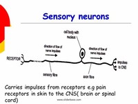
The nerve cells that make up the sensory nerves are commonly known as sensory neurons. These particular neurons are one of three different types of neurons that are found within the body. The other two neurons are known as relay neurons and motor neurons.

Trigeminal neuralgia is inflammation of the trigeminal nerve, causing intense facial pain. It is also known as tic douloureax because the intense pain can cause patients to contort their face into a grimace and cause the head to move away from the pain. The obvious movement is known as a tic.

There are three cranial nerves that innervates muscle to move the eye. The main cranial nerve that controls eye movement is occulomotor nerve (CN III). It is responsible for inferior rectus, superior rectus, medial rectus, and inferior oblique. Lateral rectus muscle is innervated by abducens nerve (CN VI).

VII facial (mixed) Sensory fibres are concerned with taste via the taste buds at the front of the tongue. Motor fibres control secretion of tears via the lacrimal glands and saliva via the sublingual salivary glandsas well as facial expressions via some of the muscles of facial expression.