Types of Xrays

A breast MRI is mainly used for women who have been diagnosed with breast cancer, to help measure the size of the cancer, look for other tumors in the breast, and to check for tumors in the opposite breast.
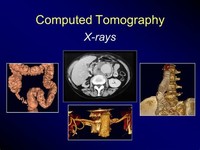
Computed Tomography (CT) Although also based on the variable absorption of x rays by different tissues, computed tomography (CT) imaging, also known as "CAT scanning" (Computerized Axial Tomography), provides a different form of imaging known as cross-sectional imaging.

Computed tomography (CT) scan, also called computerized axial tomography (CAT) scan, is used to create cross-sectional images of structures in the body. In this procedure, x-rays are taken from many different angles and processed through a computer to produce a three-dimensional (3-D) image called a tomogram.

A barium X-ray is a radiographic (X-ray) examination of the gastrointestinal (GI) tract. Barium X-rays (also called upper and lower GI series) are used to diagnose abnormalities of the GI tract, such as tumors, ulcers and other inflammatory conditions, polyps, hernias, and strictures.

Magnetic resonance imaging (MRI) is a test that uses powerful magnets, radio waves, and a computer to make detailed pictures inside your body. Your doctor can use this test to diagnose you or to see how well you've responded to treatment.

Magnetic resonance imaging can be performed on any part of the body and, unlike other imaging procedures (e.g., CT scan, x-ray), does not involve radiation. Compared to CT scan, MRI produces more detailed images of soft tissue and organs, differentiates between similar tissues more effectively, and produces less detailed images of bone.

A mammogram is a low-dose x-ray that allows specialists to look for changes in breast tissue. Screening mammograms are used to look for breast changes in women who do not appear to have breast problems.

What is an MRI? Magnetic Resonance Imaging, commonly referred to as MRI, is not an x-ray. It does not use or produce radiation. It uses a magnetic field, radio waves and a computer to produce a very clear image of the structures inside your body.

Positron emission tomography (PET) is a nuclear imaging technology (also referred to as molecular imaging) that enables visualization of metabolic processes in the body. The basics of PET imaging is that the technique detects pairs of gamma rays emitted indirectly by a positron-emitting radionuclide (also called radiopharmaceuticals, radionuclides or radiotracer).

Nuclear medicine is a branch of medical imaging that uses small amounts of radioactive material to diagnose and determine the severity of or treat a variety of diseases, including many types of cancers, heart disease, gastrointestinal, endocrine, neurological disorders and other abnormalities within the body.

Both ultrasound and X-ray technology tend to be readily or widely available, and relatively inexpensive compared to other modalities. So what is the difference between the two? Each imaging modality, whether it be X-ray, ultrasound or another, has their pros and cons.
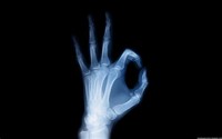
X-rays are a form of electromagnetic radiation, as are radio waves, infrared radiation, visible light, ultraviolet radiation and microwaves. One of the most common and beneficial uses of X-rays is for medical imaging. X-rays are also used in treating cancer and in exploring the cosmos.

X-rays and other radiographic tests help doctors look for cancer in different parts of the body including bones, and organs like the stomach and kidneys. X-rays are typically fast, painless, and there’s no special preparation needed.